
The University of Texas at Dallas Imaging and Histology Core facility is located in the Natural Science and Engineering Research Laboratory (NSERL) with 3090 square feet of renovated and dedicated lab space. The core houses state-of-the-art instruments and technology for on-campus researchers and external users to provide affordable support for basic and translational research in biological and interdisciplinary sciences. This core facility is operated by the Office of Research and Innovation and employs dedicated full-time technical staff to operate and maintain the equipment. The Imaging Core houses advanced imaging systems, including an electron microscope, a confocal laser scanning microscope, a high-throughput slide microscope, a multiphoton intravital imaging system, and various other optical microscopes. The Imaging Core also houses workstations equipped with advanced image analysis software such as Imaris, cellSens, and Fiji.
The Histology Core provides support for processing and embedding of tissue samples, sectioning and slide preparation, cell culture, and sample imaging.
Olympus and Zeiss Imaging Instruments
At the UTD Imaging Core, much of the equipment is part of the Olympus Discovery Center, a collaborative effort between The University of Texas at Dallas and Olympus. This collaboration established a state-of-the-art imaging core, and includes top-end Olympus imaging facilities, hands-on training from Olympus staff, an imaging summer program run cooperatively by Olympus and UTD, and regular beta testing of new Olympus imaging equipment.
Please contact ImagingCore@utdallas.edu for more information.
Histology and Cell Culture Equipment
- Leica ASP300 S: Fully enclosed tissue processor
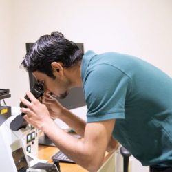
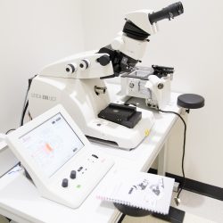
- Leica EM UC7: Ultramicrotome used for sample preparation for electron microscopy
- Leica RM2235: Manual microtome
- Leica CM1860: Clinical Cryostat
- Leica EM FC7: Cryochamber used for sample preparation for electron microscopy
- HistoCore ARCADIA C and H: Cold and hot paraffin embedding stations
- Leica EM ACE200: Carbon coater used for sample preparation for electron microscopy
- Leica EM KMR3 Glass Knifemaker: used for sample preparation for electron microscopy
- NuAire Class II, Type A2 Biological Safety Cabinet
- NuAire Direct Heat In-Vitrocell Air-Jacketed CO2 Incubators
Please contact HistologyCore@utdallas.edu for more information.
You must be logged in to post a comment.